
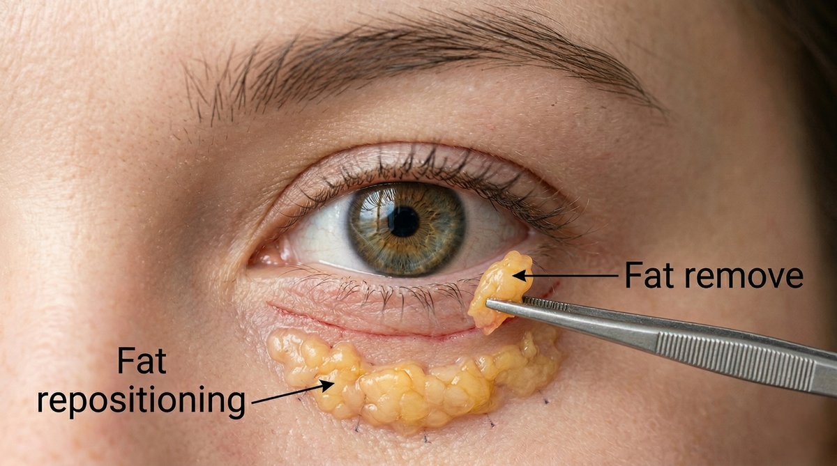
Should the Fat Under Your Eyes Be Moved or Removed?
Moved, in most eyelids. The fat that makes an under-eye bag is not surplus tissue; it is the cushion behind the eyeball that has slipped forward through a weakened membrane, and in a lower blepharoplasty it can either be cut out or laid back down over the bone into the groove beneath the bag. Cutting it out flattens the bag today and empties the eyelid for the decades that follow. Moving it flattens the bag and fills the groove with the same tissue. Removal still has a place, but as a measured trim after the fat has been moved, not as the plan.
Most patients are never told which of the two their surgeon intends to do. “We take the bags away” describes both operations equally well, and it leaves out the only thing that matters: where the fat goes. On a face that keeps losing volume every year after surgery, a lower eyelid that starts from an emptied orbit and one that starts from a redistributed orbit age in opposite directions.
The case for moving the fat rests on four things: what removal left behind during the fifty years it was standard, where the fat sits and what holds it there, what the measured comparisons between moving it and removing it found, and a judgment made on the table about how much to move and how much to trim.
Why Did Surgeons Cut the Fat Out for Decades, and Why Did They Stop?
Surgeons removed the fat because the bag looked like too much fat, and removing it worked for long enough to be convincing. Through most of the twentieth century the operation opened the septum, teased out the herniated fat and excised it, and the eyelid was flat the following week. What that eyelid looked like ten years later was not part of the follow-up.
Sam Hamra was among the first to say in print what he had seen in those later faces. In 1995 he wrote that conventional techniques removing lower eyelid fat can create a concave contour deformity that gives the eye an operated look, and proposed keeping the fat and resetting the septum instead. Five years later Robert Goldberg published the version most surgeons now recognize: in 24 patients followed for 6 to 30 months, the medial and central fat were rotated over the orbital rim into a pocket beneath the periosteum and held there with a polypropylene stitch passed out through the cheek skin and removed after 3 to 5 days.
Why an emptied eyelid looks worse with time comes down to what the rest of the orbit does. On MRI of 40 people in three age bands, Darcy and colleagues measured the soft tissue in front of the lower orbit growing from 99 to 103 to 131 square millimeters across the age groups, with fat expansion the largest single contributor, from 28 to 43 square millimeters, while the eyeball itself stayed where it was. The bone moves too. Pessa’s three-dimensional CT of 28 subjects found the orbital rim retreating relative to the cornea with age, and he warned that over-resecting orbital fat accentuates the prominent-eye appearance that aging produces on its own. An eyelid emptied of fat sits over a rim that is moving backward, and the eye reads as more prominent and more hollow at once.
How often removal produces that hollow has barely been measured. A 2025 systematic review of 36 series still records periorbital hollowness in 0 to 9.8 percent and inappropriate fat removal in 0 to 4.9 percent, as ranges between studies rather than a pooled rate. The one attempt to measure hollowing directly is small: in 39 patients who had a mean of 0.55 mL of fat removed per eyelid, Schiller and colleagues found a visibly deeper upper eyelid sulcus in 10 percent afterward, a finding that drew two published letters of disagreement. Everything else, including Hamra’s word for it, “frequent,” is the opinion of surgeons who have seen the fifteen-year-old fat-removal eyelid and did not count. I am one of them. Darcy’s own MRI paper ended by recommending that some fat excision remain part of treatment, and on that narrow point I agree: a measured trim, taken after the fat is already lying in the groove, is where excision still has a place.
What Is the Fat Under Your Eye, and Where Exactly Does It Sit?
It is orbital fat, the cushion that surrounds the eyeball, held in front by a thin fibrous sheet called the orbital septum, which fuses with the eyelid’s deeper layer 3.7 to 5.4 mm below the lower edge of the tarsal plate. A bag is not new fat. It is the same fat pressing forward through a septum that has thinned, and it stays in front of the bone regardless of sleep, salt or allergies.

Textbooks divide the lower orbital fat into three pads: medial, closest to the nose; central, the largest; and lateral, toward the temple. Between the medial and central pads runs the inferior oblique muscle, one of the muscles that rotate the eye, and the fascia of that muscle is the partition. The tidy picture is less reliable than the drawings suggest. When 50 cadavers were microdissected, the classic three-pad pattern was present in only 36 percent; 20 percent had two pads and 44 percent had a double-convexity arrangement. A surgeon who assumes the map instead of finding the muscle is working from a diagram that fits one orbit in three. The muscle originates on the orbital floor 5.14 mm behind the rim and lies in the direct path of a transconjunctival dissection, and in 56 Japanese cadaver orbits its nerve entered the muscle a mean of 3.4 mm below the inferior orbital rim.
Beneath the fat is the line that makes the shadow. In 48 cadaveric hemifaces, Wong, Hsieh and Mendelson described a true ligament running from the bone to the skin, from the medial canthal tendon to about the medial pupil line, and continuing outward as the orbicularis retaining ligament. Micro-CT of 22 cadaver specimens shows that ligament as a multilayered fibroelastic meshwork rather than a cord, splitting laterally into two layers 4 to 8 mm apart. Fat that is to fill the groove has to cross that line, which is why releasing it is part of the operation and not an optional step.
East Asian orbits change the arithmetic. On three-dimensional CT of 224 Japanese adults, Kato and Okuda found the infraorbital rim holding at 7.8 mm behind the front of the cornea in every age band from the twenties to the seventies, while the front edge of the orbital fat advanced from 4.4 mm to 2.4 mm behind the cornea. The bag comes forward; the rim does not retreat. A CT study of 107 Koreans found no significant enlargement of the bony orbital aperture with age in either sex, unlike the widening described in 60 white subjects that Western articles cite reflexively. And CT volumetry of 130 Koreans found orbital fat volume rising until about age 60 and then falling. Put together: a Korean under-eye bag is usually fat advancing over a rim that is standing still, which is the anatomy repositioning was designed for. The volumetry authors read their own curve as a case for conservative excision, which is what a trim measured after the fat has been repositioned amounts to.
Fat Removal, Fat Repositioning, or Removal With Fat Grafting: How Do the Three Compare?
Three operations handle the same fat three ways: cut it out and discard it, move it on its own blood supply, or cut it out and put it back as a graft. The second and third both keep the fat in the eyelid, which puts them in one family against the first. Direct comparisons between the three are few, and the ones that exist differ widely in how long they followed patients.
The cleanest head-to-head is a 2019 study of 60 patients and 120 eyelids from Goldberg’s group, comparing fat excision with fat transposition at a mean of 5.6 months. Excision left a shorter measured lower eyelid, 13.5 mm against 16.1 mm. Transposition erased the nasojugal fold far more completely, an effacement score of 1.5 against 0.48. The authors read the pair of results as a trade-off a surgeon balances rather than a winner. Since the fold is what casts the shadow, and the shadow is what the patient came in about, the second number is the one I weigh.
Between the two ways of keeping the fat, the differences are small. A Swedish series of 339 lower blepharoplasties compared transposition of the fat with cutting it into segments and grafting it back, at a mean follow-up of 12.9 months: complications 4 percent, revisions 9 percent, no significant difference between the methods. The largest recent cohort of the free-graft version, 200 consecutive extended transconjunctival lower blepharoplasties followed for a mean of 22 months, reported 99.0 percent satisfaction, no lower eyelid malposition, no ectropion, fat necrosis in 3.5 percent and revision in 1.0 percent, with no patient receiving a canthopexy, canthoplasty or skin pinch. Those are among the strongest safety figures published, and they belong to an operation that kept every gram of fat.
Removal followed by grafting has its own Korean series. In 229 patients in Korea, the fat was excised, minced and injected back; 97.8 percent were satisfied, and graded fat prolapse fell from 1.94 to 0.07. That satisfaction figure rests on short follow-up: the mean was 6.45 months, and two-thirds of the patients were assessed at one to two months. Fine wrinkling appeared in 60.7 percent and infraorbital hollowness in 3.0 percent, and the authors used the series to argue that repositioning risks scarring the eyelid’s middle layer. A second Korean comparison, 120 patients treated either by pedicled fat redraping or by septal reset, found septal reset gave better grade improvement in the deeper grooves, with no major complications in either group. More than one way of keeping the fat satisfies patients. Removing it and discarding it is the one approach none of these series defends.
Where the moved fat is laid has been compared exactly once. In 2013 Yoo, Peng and Massry set 20 subperiosteal against 20 supraperiosteal transpositions and found no statistically significant difference in blinded aesthetic outcome; the supraperiosteal operation was faster, and in the authors’ impression bruised more. Thirteen years later no second comparison has been published, so the plane remains the surgeon’s choice rather than a question the evidence settles.

Fat Removal vs. Fat Repositioning vs. Removal With Grafting at a Glance
| Fat removal alone | Fat repositioning (transposition or same-site free graft) | Removal with fat grafting | |
|---|---|---|---|
| What happens to the fat | Excised and discarded | Kept in the eyelid and laid over the rim into the groove | Excised, minced, injected back |
| The bag | Corrected | Corrected | Corrected |
| The groove below it | Untouched, often deeper as the face ages | Filled with the patient’s own fat | Filled with grafted fat, part of which is resorbed |
| Orbital volume kept | No | Yes | Partly |
| Hollow eyes years later | The recognized risk; hollowness 0 to 9.8 percent across 36 series of mixed technique | Not the pattern of this operation | Hollowness 3.0 percent within months in a 229-case series |
| Characteristic failure | Under-excision 7.4 percent in a 122-case series; hollowing | Residual bag or lump; revision 1.0 to 9 percent across series; 31 of 183 needed a second volume procedure when grafting was added | Fine wrinkling 60.7 percent; over- or under-correction |
| Lower lid position | Route-dependent | 0 malposition in 200 transconjunctival cases | Transient ectropion 0.4 percent in 229 cases |
| Scar | None if transconjunctival | None if transconjunctival | None if transconjunctival |
| Suits | Very few eyelids; a measured trim as part of repositioning | A bag with a groove beneath it, most patients | Surgeons trained in it; short-term results comparable to repositioning |
How I Reposition the Fat, and When I Trim Some of It
Repositioning is my default, and removal happens only after the fat is already lying in the groove and the eyelid is still too full. The order is fixed, from the examination to the last knot, and the trim comes at the end of it or not at all.
I choose the incision by the skin, not by the fat. When fat has come forward and the groove has appeared but the skin has not loosened, which in my patients is most often between the mid-thirties and the late forties, I go through the conjunctiva and leave the skin untouched. A small excess is handled with a narrow pinch of skin under the lashes. When the case is about loose, redundant skin, usually later in life, I use the skin incision, because an operation done from inside the eyelid cannot tighten what it never touches. That incision sits 1 to 2 mm below the lash line, and I raise a skin-muscle flap rather than a skin-only flap: skin and orbicularis come up together to 4 to 5 mm below the lash line to preserve a cuff of pretarsal orbicularis, the muscle is then divided where it inserts on the tarsal plate, and the dissection continues in the plane deep to the orbicularis and superficial to the still-closed orbital septum.

I test the eyelid before I plan the fat. Two bedside tests decide whether the eyelid can hold repositioned fat: the snap-back test, and the distraction test with the thresholds I work to, 3 to 5 mm normal, 6 to 7 mm significant laxity, and 10 mm or more meaning I plan a horizontal shortening of the lid. Both tests, and the reasons behind the numbers, are described in tear trough filler versus lower blepharoplasty. A lax lid with fat stitched beneath it is a retraction waiting to happen.

I find the inferior oblique before I touch the medial fat. Because the three-pad map fits only one orbit in three, I do not assume where the muscle is. It is identified between the medial and central fat before either pad is mobilized. This is the muscle whose injury produces double vision, and the one step in the operation that is never hurried.
I release the ligament and lay the fat on the periosteum. After the septum is opened, the tear trough ligament is released from the bone so that the fat can cross it, and I dissect the pocket over the rim in the supraperiosteal plane, above the bone’s covering rather than beneath it. The one comparison of the two planes found no difference in the result, so the choice comes down to what the fat can be fixed to; I keep the periosteum intact as the bed and stitch to it. Into that pocket I bring the medial, the central and the lateral fat.
I fix the fat from inside with a Chang’s needle. The fat has to stay where it is put while it heals. The older method passed sutures out through the cheek skin and tied them over bolsters for several days, which is what Goldberg described in 2000. I use the internal fixation developed in Korea and sold as EZ-Tcon: a short, curved, double-ended needle that passes out through the cheek tissue and back in, catching the fat and the tissue beneath it, so the fat is drawn snugly into the pocket and held there without a stitch on the skin. In the 545-patient comparison of externalized sutures against this internal fixation, complications fell from 11.4 to 1.5 percent, visible suture marks on the eyelid from 8.1 percent to none, and reoperation from 2.8 to 0.6 percent, at a median follow-up of 70 days. My suture is 5-0 PDS, absorbable, tied in three to four knots to the periosteum about 4 to 5 mm below the nasojugal groove at the upper edge of the cheek fat, the level at which the transposed fat erases the groove from below.

Then, and only then, I decide whether to trim. With the fat down and fixed, I look at the eyelid. If it is level, nothing is removed. If it still looks too full, because the bag was large and the groove shallow, I trim a measured amount rather than forcing all of it into a groove that cannot hold it. That trim is the only fat removal in my lower blepharoplasty, and it is decided by what the eyelid looks like with the fat in its new place, never by what it looked like before the fat was moved. The amounts involved are small, a fraction of what a removal-first operation takes.
A thin face gets fat added, not removed. When there is little orbital fat to move and a deep groove to fill, repositioning alone can leave the eyelid looking as though it will hollow once the swelling settles. In that case I add a small amount of micro fat grafting in the same operation. Otherwise I wait about three months for the contour to settle and add fat only if the patient still wants more. How much of a graft survives is not fully predictable, which is covered in fat graft survival.
If filler is already in the groove, it comes out first. I dissolve it with hyaluronidase at least one to two weeks before surgery and re-examine before fixing the date, because I cannot plan how much fat to move over a groove I cannot see. The dose and the waiting are described in the overfilled face.
Sedation differs from my upper eyelid surgery. For lower blepharoplasty I use propofol sedation with local anesthesia, including an infraorbital nerve block, rather than the midazolam and ketamine I use for double eyelid surgery. The lower eyelid does not need to be checked in motion the way a crease does, and a still, comfortable patient matters more here.
Which Lower Eyelid Is Yours? Five Profiles
Five lower eyelids ask for the same thing and leave with different plans. The variable is not age. It is how much fat there is to move and how much groove there is to fill.
A bag with a groove beneath it and skin that is still tight. The commonest eyelid in my practice, most often between the mid-thirties and late forties. All of the fat is moved, nothing is trimmed, and the skin is not touched.
A large bag over a shallow groove, in a full cheek. Recognizable because the bulge stands well above the rim while the groove beneath it is faint. More fat than the groove can hold, so this is the one profile in which some fat leaves the eyelid, and it leaves last.
A small bag over a deep groove, in a thin face. Recognizable because the shadow is long and the bulge is slight. Not enough fat to fill the valley, so this is the profile that gets fat added rather than taken away.
A bag in the late twenties with a Korean or East Asian eyelid. Fat forward of a rim that has not retreated and will not. This is anatomy rather than aging, and it is treated the same way as the first profile, with the skin left alone.
A hollow from a fat-removal operation years ago. The fat that could have filled the groove is gone, and the eyelid is being revised, not treated for the first time. The tool is fat grafting. A meta-analysis of 39 studies and 4,046 lower-eyelid fat grafting cases puts satisfaction at 90.9 percent and complications at 7.9 percent, and in a 183-patient Asian series combining repositioning with grafting, 11 patients were over-corrected and 20 under-corrected, 31 in all who needed a second volume procedure. Scar from the first operation changes the planes and makes graft survival less predictable, which is why this eyelid gets the most conservative plan and the most explicit warning.
Loose skin cuts across all five profiles. When the skin has loosened, most often from the fifties onward, the same fat operation is done through a skin incision with the excess removed, and the canthus is supported when the distraction test calls for it.
What Does Recovery Look Like After Fat Repositioning?
Judge the eyelid at three months. Before that it is swollen, then bruised, then subtly full as the moved fat softens, and none of those stages is the result.
After transconjunctival surgery there are no skin stitches to remove, only a wound check at day five to seven; after a skin incision the sutures come out on day five to seven. Bruising fades over one to two weeks, most patients are presentable with light makeup at about ten days, and the final contour arrives at two to three months as the repositioned fat settles. In the 200-case transconjunctival series, chemosis, a jelly-like swelling of the conjunctiva, occurred in 3.0 percent overall and in none of the patients who had the lower eyelid operation alone. A transient foreign-body feeling in the eye for two or three days is common and expected. Patients traveling for the operation should plan to stay in Korea roughly a week, set out procedure by procedure in how long to stay in Korea after plastic surgery.
One symptom is never waited on. Severe pain with pressure behind the eye in the first hours is a bleeding emergency, which is why patients are told to stay within reach of the clinic on the first night.
Surgeon’s Insight
Patients ask me whether I will remove the fat or move it, as if those were two schools and I belonged to one. The honest answer is that I move all of it, look at the eyelid, and then decide whether any comes out. Order matters more than philosophy here. Fat trimmed before it has been moved is trimmed against the bag; fat trimmed after it has been moved is trimmed against the groove, and the groove is the thing the patient is actually looking at. Most days the answer, once the fat is down, is that nothing needs to come out at all.
What Can Go Wrong When Fat Is Moved, and When It Is Removed?
Repositioning fails by doing too little; removal fails by doing too much. Both are visible, but only one of them is reversible by adding tissue, and that asymmetry decides which of the two errors is worth risking.
When fat is removed. Hollowing is the recognized late outcome, reported in 0 to 9.8 percent of patients across 36 series, and the early one is under-correction: in a 122-case transconjunctival series, the main complication was under-excision of fat in 7.4 percent, because a surgeon who fears the hollow removes too little and leaves the bag. The Korean removal-and-grafting series recorded fine wrinkling in 60.7 percent and hollowness in 3.0 percent within months, and hollowing that appears years later is corrected only by grafting, which carries a 7.9 percent complication rate in pooled lower-eyelid data.
When fat is moved. In a 150-case series of transconjunctival fat repositioning, 141 patients were very satisfied; the recorded problems included a residual lateral fat pad in 3, temporary medial ectropion in 2, conjunctival granuloma in 2, retraction in 1, and three temporary inferior oblique palsies, all of which resolved. A pooled review of 24 cohorts and 3,623 patients recorded, after the transconjunctival route, delayed swelling, induration or granuloma in 3 percent, under-correction in 0.8 percent and hematoma in 0.3 percent, with ectropion in 1 of 2,266 patients against 3 of 182 after skin incisions, a difference that did not reach significance. In the 200-case series, scarring of the eyelid’s middle layer occurred in 1.0 percent and lower eyelid malposition in none. Releasing the retaining ligament, which the fat must cross, has been reported to prolong swelling and to raise the chance of ectropion in one 53-patient comparison, while the surgeons who developed the extended release report zero malposition in 200 cases; the literature is split, and the lid laxity tests are how I decide how much the eyelid can tolerate. Adding a skin pinch does not lower the lid: in 45 patients measured with and without it, the lower eyelid position changed by 0.05 mm either way.
Double vision, from either operation. Injury to the inferior oblique muscle is the complication specific to this part of the orbit. Across the 36 series in the 2025 review, no case of diplopia or inferior oblique injury was recorded, and the 3,623-patient pooled review found diplopia in 1 patient after the transconjunctival route; the 150-case repositioning series recorded three temporary palsies that all resolved. Registry data show the risk is real but small: among 368,623 blepharoplasty patients in the IRIS Registry, strabismus surgery within three years was about two and a half times as likely after lower as after upper blepharoplasty, 0.15 against 0.06 percent, a difference that is real but small in absolute terms and not a measure of how often the operation caused it. When 371 oculoplastic surgeons were surveyed, 23.2 percent had seen at least one case of persistent double vision after lower blepharoplasty in their careers, the inferior oblique was implicated in 61 percent of those cases, and 73 percent resolved completely. That is a rate per surgeon, not per patient. It is also the reason the muscle is identified before the medial fat is touched.
Bleeding. Rare, early, and the one emergency. Blood collecting behind the eye threatens vision through pressure on the optic nerve, which is why that pain is the one symptom a patient is told to act on at once rather than sleep on.
The Question to Ask: What Happens to the Fat?
Do not ask whether you need lower blepharoplasty. Ask this: what happens to the fat?
If the answer is that it will be removed, ask what will fill the groove beneath it, this year and in fifteen years. If the answer is that it will be moved, ask where it will be fixed and how, and whether any will be trimmed after it is in place. A surgeon who answers with the plane, the fixation, and the rule for trimming has told you what your eyelid will look like at fifty, which is the only time horizon on which this operation is judged. Under either plan the bag is gone at one week. Everything that happens after that is the difference between the two.
Written by Dr. Yongwoo Lee, board-certified Korean plastic surgery specialist in facial anatomy and aesthetic procedures at VIP Plastic Surgery, South Korea.
Frequently Asked Questions About Fat Repositioning and Fat Removal
What is the difference between fat repositioning and fat removal in lower blepharoplasty?
Both operations open the septum and reach the orbital fat that makes the bag. Removal excises that fat and discards it. Repositioning keeps it on its blood supply, lays it down over the orbital rim into the groove beneath the bag, and fixes it there, so one tissue corrects both the bulge and the hollow. In a 120-eyelid comparison, transposition erased the nasojugal fold far more completely than excision, 1.5 against 0.48 on the effacement scale used.
Why does fat removal cause hollow eyes years later?
Because the face keeps changing after surgery from a starting point that has already been emptied. MRI across three age bands shows periocular fat increasing with age, and separate three-dimensional CT shows the orbital rim retreating relative to the cornea in Western subjects, so an eyelid stripped of fat sits over a receding bone and reads as both prominent and sunken. In East Asian orbits the rim holds its position and the bag is fat advancing over it; either way, removing the fat removes the only tissue available to fill the groove. Series report periorbital hollowness in 0 to 9.8 percent of patients, and a dedicated study found a visibly deeper sulcus in 10 percent after a mean removal of only 0.55 mL per eyelid.
Is fat repositioning better than fat removal?
For an eyelid with a bag and a groove beneath it, yes, because it treats both with the patient’s own tissue and does not empty the orbit. Removal alone is defended by none of the recent comparative series. Where it still belongs is as a measured trim after the fat has been moved, in the eyelid whose bag is larger than the groove can hold.
Does any fat get removed during fat repositioning?
Sometimes, and only after the fat is in its new place. In my operation all three fat pads are moved into the groove and fixed first; if the eyelid still looks too full, a small measured amount is trimmed. The decision is made with the fat down, not before, because the same fat that is excess against the bag may be exactly enough against the groove.
Where is the fat placed, and how is it held there?
Over the orbital rim into a pocket on the periosteum, after the tear trough ligament has been released so the fat can cross it. I hold it with an internal fixation using a Chang’s needle, sold as EZ-Tcon, and an absorbable 5-0 PDS suture tied in three to four knots to the periosteum about 4 to 5 mm below the nasojugal groove. In a 545-patient comparison, internal fixation reduced complications from 11.4 to 1.5 percent and eliminated the suture marks left by the older method of stitching out through the cheek skin.
Subperiosteal or supraperiosteal: does the plane matter?
The only comparison, 20 patients per plane at a mean of 10 months, found no statistically significant difference in blinded aesthetic outcome, and no second comparison has been published since 2013. I use the supraperiosteal plane and stitch to the intact periosteum beneath it.
Does fat repositioning leave a scar?
Not on the skin when done through the conjunctiva, which is the route for patients whose skin is still elastic. When loose skin also has to be removed, the incision runs just under the lashes and fades into the lash shadow. In pooled data from 3,623 patients, ectropion followed 1 of 2,266 transconjunctival operations and 3 of 182 skin-incision operations.
How long do the results of fat repositioning last?
The correction is structural and the eyelid ages normally around it. In a 200-case transconjunctival cohort followed for a mean of 22 months and up to 95 months, revision was needed in 1.0 percent; in a 339-case Swedish series, revisions ran at 9 percent. No series with a five-year mean follow-up has been published. Past that point the fat stays where it is fixed, and the face continues to change around it without the emptied starting point that removal leaves.
Can the repositioned fat be reabsorbed?
The fat is moved on its own blood supply, so it is living tissue rather than a graft, and no series has quantified how much of a transposed pedicle resorbs. What patients notice more often is the moved fat softening over two to three months, which is settling, not disappearance. Grafted fat behaves differently: a proportion of any injected graft is resorbed, which is why grafting plans are conservative.
Can hollow eyes from an old fat-removal blepharoplasty be fixed?
Yes, with fat grafting, and with realistic expectations. Meta-analysis of 4,046 lower-eyelid fat grafting cases puts satisfaction at 90.9 percent and complications at 7.9 percent, and in a 183-patient series 31 needed a second procedure for over- or under-correction. Scar from the first operation alters the tissue planes and makes graft survival less predictable, so revision is planned in stages rather than corrected in one sitting.
Can lower blepharoplasty cause double vision?
Rarely. The inferior oblique muscle runs between the medial and central fat pads and can be injured during dissection. Across 36 published series no case of inferior oblique injury was recorded; a 150-case series reported 3 temporary palsies, all resolved; and in a registry of 368,623 blepharoplasty patients, strabismus surgery within three years was about two and a half times as likely after lower as after upper blepharoplasty, 0.15 against 0.06 percent, small in absolute terms. Identifying the muscle before touching the medial fat is the safeguard.
Am I too young for fat repositioning if I am in my twenties?
Age is not the criterion; fat position is. In 224 Japanese adults on CT, the orbital rim sat 7.8 mm behind the cornea in every age band from the twenties to the seventies while the fat advanced steadily, so an East Asian bag in the late twenties is structural rather than age-related. If the fat is in front of the bone, the operation is the same; what age changes is whether the skin also needs tightening.
What is the recovery time after fat repositioning blepharoplasty?
No skin stitches after the transconjunctival route, a wound check at day five to seven, bruising for one to two weeks, light makeup at about ten days, and the final contour at two to three months as the moved fat softens. Chemosis occurred in 3.0 percent of a 200-case series and in none of the patients who had the lower eyelid operated on alone.
Should I get tear trough filler instead of surgery?
Only if there is a groove and no bag. Filler raises the floor of the groove and cannot move fat that has come forward; a bag under filler keeps growing beneath the gel. How to tell a bag from a hollow in a mirror, and what five-year follow-up on under-eye filler shows, is set out in tear trough filler versus lower blepharoplasty.
This article is for informational purposes only and does not constitute medical advice. Always consult with a qualified medical professional before making any decisions about surgical or non-surgical procedures.