
How Common Is Facelift Nerve Damage, and Can It Be Permanent?
Facelift nerve damage is uncommon and usually temporary, and it has been counted. In a 2026 meta-analysis of the deep plane facelift covering 45 studies and 10,784 patients, temporary facial nerve injury was 1.2 percent and permanent injury appeared in zero cases, meaning none reported across 45 retrospective series rather than impossible.
Clinic pages on deep plane facelift nerve damage answer with “rare and temporary,” which could mean one in ten or one in a thousand. A meta-analysis across SMAS techniques found that risk of permanent injury did not differ, and that the highest temporary rates belonged to high lateral SMAS at 1.85 percent and composite rhytidectomy at 1.52 percent. That meta-analysis reports no temporary rate for the deep plane itself.
Baker and Conley’s 1979 paper put injury under one percent, with spontaneous return of function in more than 80 percent within six months. Those are 1979 figures rather than modern deep plane numbers. A 2025 review of 20 studies reports 0.5 to 5 percent across designs that cannot be compared, and no midpoint of that range is published.
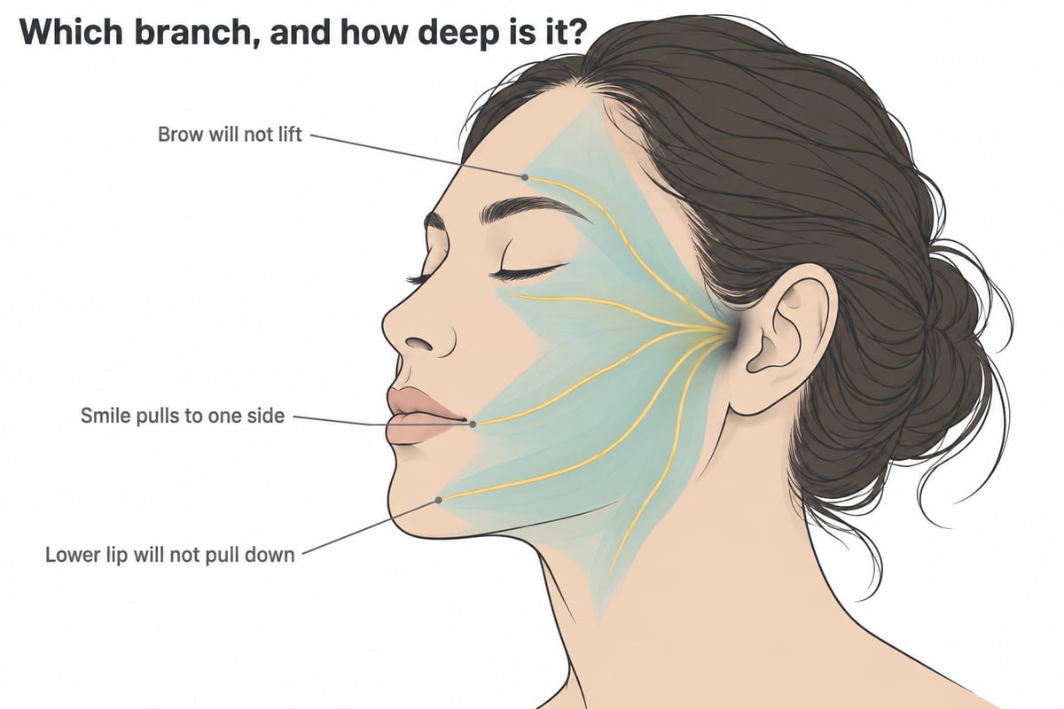
Which Branch, and How Deep Is It Where the Surgeon Is Dissecting?
Risk turns on one relationship: how deep a branch runs at the exact point the dissection has reached. Asking whether a facelift is safe for the nerve gets a marketing answer, while asking how deep the nearest branch lies where the surgeon is working gets a surgical one.
A 2020 review of facial nerve danger zones states that branching patterns of the marginal and cervical branches are variable in two dimensions, while in three their position and depth are constant and predictable. Danger zones, in its words, are where the facial nerve sits superficial and adjacent to the planes of dissection used in facelifts.
Memorizing a single line fails most plainly in the temple. In 42 hemifacial cadaver dissections, temporal branches crossing the zygomatic arch numbered one to three, the nearest 20.62 plus or minus 3.84 mm from the tragus on the right and 21.33 plus or minus 3.10 mm on the left.
Deep plane facelift safety cuts both ways: sub-SMAS dissection is protective where branches run under a fascial floor and closest to one where the retaining ligaments are divided, as in what a deep plane facelift actually releases.
Which Facial Nerve Branches Does a Facelift Put at Risk, and Why?
Five branch groups leave the parotid gland: temporal (frontal), zygomatic, buccal, marginal mandibular and cervical. A facelift crosses all five territories at five different depths.

The frontal branch, over the zygomatic arch
Patients who cannot raise one eyebrow after a facelift are describing this branch, and the forehead on that side goes smooth with it. Fear attaches here because a brow that will not lift shows in every expression.
Anatomy over the arch is not what most textbooks taught. In 18 fresh-frozen cadaver hemifaces, the authors set the common teaching that the frontal branch travels within the SMAS at the arch against what they found: in all dissections it lay within the innominate fascia, crossing into the superficial temporal fascia 1.5 to 3.0 cm above the arch and 0.9 to 1.4 cm behind the lateral orbital rim.

A second cadaver study puts the transition lower. Across 36 hemifacial fresh specimens, the branch became intra-SMAS at a mean 9.61 plus or minus 5.08 mm above the arch and 12.19 plus or minus 4.77 mm behind Pitanguy’s line. Its authors call that a caution zone rather than a safe margin, and the 5.08 mm standard deviation is why. The two studies disagree, and a midpoint between them is a number neither one reports.
Innominate fascia is that paper’s own term and is distinct from the SMAS. A branch lying deep to the SMAS is what allows the SMAS to be divided at or above the superior border of the arch. Brow work above it has its own fixation question.
The zygomatic and buccal branches, at the retaining ligaments
A crooked smile after a facelift usually belongs here, alongside a cheek that will not lift. Upper zygomatic branches add incomplete eye closure to that picture.
Depths at the ligaments were measured in 22 cadaver hemifaces studying the zygomatic branch against the retaining ligaments. Main zygomatic and upper masseteric ligaments sat a mean 44.91 plus or minus 9.72 mm and 46.35 plus or minus 8.34 mm from the tragus. An upper zygomatic branch passed between them, always deep at 4.07 plus or minus 1.29 mm in the sub-SMAS plane. An inferior branch ran below the upper masseteric ligament or pierced its margin in 54 percent of cases, lying at 1.41 plus or minus 0.95 mm and visible just distal to it.
Those authors concluded that the ligaments create a safe passage between them through which a zygomatic branch passes deep, and the danger sits inferomedial to the upper masseteric ligament, where that branch becomes superficial and vulnerable. Ligament release, the defining maneuver of the extended deep plane, happens there.
Those depths belong to this plane alone, and ligaments vary between faces. A 2021 review of short-term facelift complications calls the buccal branch the most commonly injured yet often unnoticed because of cross-innervation. That statement is review-level, and no primary study behind it reports a rate.
The marginal mandibular and cervical branches, and the lower lip that is not paralyzed
Lower lip weakness after a facelift gets described precisely: the lip will not pull down on one side, fewer lower teeth show, and the mouth looks crooked in speech. Two different injuries produce that appearance.
Numbers come from a single surgeon’s 2002-case SMAS-platysma experience. Pseudoparalysis of the marginal mandibular nerve, caused by cervical branch injury, occurred in 34 of 2002 facelifts, 1.7 percent, with full recovery in 100 percent within 3 weeks to 6 months. True marginal mandibular injury occurred in 1 of 2002, 0.05 percent, a figure that appears in that paper’s body and not in its abstract. Both come from a series running 1977 to 2001, not a deep plane one.
Bedside distinction is simple enough to do in a mirror. Cervical branch injury can be told apart from marginal mandibular injury because the patient can still evert the lower lip, the mentalis muscle still working.

Naming came first, in a 1979 paper on the cervical branch describing a patient with a full denture smile who could no longer retrude the corner of the mouth after a platysmal lift. Depth came later: in 55 cadaver heads examined in 2023, the cervical branch trunk left the parotid capsule an average of 5 mm deeper than the platysma, SD 1.3 mm, range 3 to 7 mm, and the same authors observed lip depressor weakness recovering within 12 weeks. Neck work is sorted in neck liposuction versus a neck lift.
Why the sub-SMAS plane is the argument, in both directions
Sub-SMAS dissection is at once the most protected and most exposed thing a facelift does. Over the lower masseter it runs above a fascial floor with every motor branch beneath, and at the ligaments it runs onto one.
The protected half was mapped in 16 fresh cadavers plus several hundred rhytidectomies by Bryan Mendelson and colleagues. Their premasseter space is rhomboidal and 40 to 50 mm across, with the masseter fascia lining its floor and facial nerve branches passing under that fascia. Dissection there is bloodless and safe, in their words, because every branch lies outside the space, and they add that the SMAS incision should sit forward of the traditional preauricular location to be over it. That claim covers one bounded space rather than the midface.
Why Do Published Facelift Nerve Injury Rates Disagree So Much?
Published rates for the same complication run from about 1 percent to more than 12 percent, and that spread comes from study design rather than from the surgery.
A prospective study that examines every patient finds far more transient weakness than a retrospective chart review. In a prospective cohort of 166 primary facelift patients, transient weakness occurred in 12.4 percent of deep plane and 11.1 percent of SMAS plication cases, p = 0.70, with median recovery at 30 versus 25.5 days, p = 0.65. Pooled retrospective series report about one patient in a hundred. Both are honest: one counts every flicker at a scheduled examination, the other what a chart recorded.
| Branch | Plane relationship | Deficit | Frequency (source) | Recovery |
|---|---|---|---|---|
| Frontal (temporal) | Innominate fascia, deep to SMAS at the arch; intra-SMAS 9.6 ± 5.1 mm above it in one study, 1.5 to 3.0 cm in another (Pankratz 2020, Agarwal 2010) | Brow will not lift, forehead smooth on one side | Under 1 percent at review level; a 0.1 percent permanent figure exists only as a 2000 ASPS survey inside a 2021 review | No verified per-branch figure |
| Zygomatic | 4.07 ± 1.29 mm deep between the zygomatic and upper masseteric ligaments, 1.41 ± 0.95 mm just inferomedial to the latter (Alghoul 2013) | Uneven smile, weak cheek, incomplete eye closure | No branch-specific rate verified | Resolves within 3 to 4 months (Sinclair 2021) |
| Buccal | Under the masseter fascia in the premasseter space (Mendelson 2008) | Weak cheek, food pocketing, often unnoticed | Commonest at review level, often unnoticed; no verified number | Resolves within 3 to 4 months (Sinclair 2021) |
| Marginal mandibular | Depth constant in three dimensions, position variable in two (Stuzin · Rohrich 2020) | Lip will not pull down, cannot evert | 1 of 2002, SMAS-platysma series (Daane · Owsley 2003) | No verified figure |
| Cervical (pseudoparalysis) | Trunk about 5 mm deeper than the platysma, SD 1.3, range 3 to 7 mm (Minelli 2023) | Same crooked lip, but it everts because the mentalis works | 34 of 2002, 1.7 percent (Daane · Owsley 2003) | Full recovery in 100 percent, 3 weeks to 6 months; lip depressor weakness by 12 weeks (Minelli 2023) |
| All branches, deep plane pooled | not applicable | not applicable | Temporary 1.2 percent, permanent 0 reported in 45 studies, 10,784 patients (Koroma 2026) | Neurapraxia is 80 to 90 percent of injuries, with 70 percent fully recovered by six months (Pourhoseini 2025) |
Most branch-level rates have never been measured, which is why several cells carry no number.
Single-surgeon series report their own figures. A 153-patient minimal access deep plane extended series reported 1.3 percent temporary injury, retrospective, mean follow-up 12.7 months. A 2026 series of 240 extended deep plane facelifts reported transient dysfunction in 14 patients, 5.8 percent, its commonest complication. A 2026 comparison in 70 patients saw no permanent injuries, on a denominator too small for a 1 percent event. A 2025 meta-analysis of 47 studies found rates similar between planes and published no pooled percentage.
Every one of those comparisons is deep plane against other SMAS operations. No source compares it with a mini or skin-only lift for nerve injury, and a shorter incision is a design choice rather than a different operation.
How Long Does Facial Nerve Recovery After a Facelift Take?
Most weakness after a facelift is neurapraxia, the mildest grade of nerve injury, and weakness that outlasts the local anesthetic is expected to settle within 3 to 4 months. Timing depends on which rung the injury sits on, and the first rung is not an injury at all: asymmetry on the day of surgery often reflects residual local anesthetic, which dissipates in hours.
Grades come from Seddon’s framework, set out in the StatPearls chapter on peripheral nerve injury. Neurapraxia brings transient weakness with complete spontaneous recovery within days to weeks. Axonotmesis adds loss of motor and sensory function with atrophy, where recovery takes longer but remains possible, and neurotmesis is complete disruption of axons and connective tissue, where spontaneous regeneration is impossible. Axons regenerate at about 1 mm per day, range 0.5 to 3 mm, which is general nerve physiology rather than a facelift figure.

Pooled across 20 heterogeneous studies, neurapraxia accounts for 80 to 90 percent of cases, 70 percent recover fully within six months with corticosteroids and physiotherapy, and about 10 percent are left with persistent deficits. For asymmetry that persists, the same review describes chemodenervation of the opposite side with botulinum toxin as a holding measure. No verified source gives a dose for it.
For an international patient the practical question is how much of the recovery passes before anyone at home sees her, and the answer is in how long to stay in Korea.
How I Dissect Around the Facial Nerve, and Where the Plane Comes Closest to a Branch
Most of the facelifts I perform are deep plane operations, and the facial nerve is part of why that technique earns its difficulty rather than a reason to avoid it.
Why I use the deep plane, and what that means for the nerve
My reason for choosing this plane is mechanical rather than neurologic: a tightened SMAS pulls against tension that was never released, and a face freed only in part keeps its old tether. Nerve safety in that plane is the anatomists’ finding rather than mine.
Where the release comes closest to a branch
Every zygomatic ligament I can reach comes down, the masseteric ligaments along the front border of the masseter are divided, and I keep going until the tissue slides freely under my hand: medially past the nasolabial fold, just superficial to the zygomaticus major and minor, until the melo fat pad is mobilized.
That territory is the one the 22-hemiface ligament study measured, and it is where the published margin between plane and motor branch is thinnest.
Asian anatomy shrinks the margin, and what that changes
Dissecting beneath the SMAS in my patients carries one characteristic difficulty: the boundary between the SMAS and the zygomaticus major and minor is frequently indistinct. That spot is also the most dangerous, since the correct plane lies immediately over those muscles and their nerve supply.
Asian tissue, skin and SMAS alike, is markedly less elastic, so an incomplete release has no give to borrow from and has to be carried further rather than stopped short. A face that emptied rather than fell changes the calculation again, the subject of a face that lost volume rather than position.
The anatomy course, and what it does and does not prove
I have completed the Mendelson Advanced Facial Anatomy Course (MAFAC), a cadaver dissection course in facial anatomy. It is named for Bryan Mendelson, the author of the premasseter space study. Its own site records 32 courses since 2009, in a two-day format, for practicing plastic surgeons and trainees.
Dissection anatomy matters for the reason the danger-zone review gives: a branch pattern cannot be memorized, because it varies, while depth holds still and is learned on a specimen rather than off a diagram. Attending a course teaches that depth on a specimen and nothing beyond it. No published study links attendance at a cadaver course to lower facelift complication rates, and what the evidence does connect is experience to lower overall rates.
Which One Are You
Timing and finding sort these patients, not age.
The patient on day one whose smile is uneven. Residual local anesthetic explains most of these, and it dissipates in hours.
The patient at week three whose lower lip will not pull down. Eversion splits this in two: a lip that rolls outward points to cervical branch pseudoparalysis, which recovered fully in every published case, while one that will not points to the marginal mandibular branch.
The patient at month two whose brow will not lift. Frontal branch weakness shows in every expression, so what matters is the injury grade and what 1 mm per day implies.
The patient who has read that deep plane is more dangerous. Permanent injury risk did not differ among SMAS techniques, and the highest temporary rates belonged to high lateral SMAS and composite rhytidectomy.
The patient considering a second facelift. Scarred planes and a thinner SMAS are the documented finding in revision surgery, and filler complicates it further, as in an overfilled face.
The patient asking whether a smaller operation is safer. No source compares deep plane with a mini or skin-only lift, and a shorter scar moves no branch.
What Should I Do if Something Is Not Moving After My Facelift?
Check three things at home, then call rather than wait: roll the lower lip outward to see whether it everts, raise both eyebrows, and close both eyes gently to see whether the lashes disappear. On day zero this usually settles within hours, and anything still present after that gets reported rather than watched.
One finding is urgent: an eye that will not close fully leaves the cornea exposed, which is a reason to be seen the same day. Otherwise the published treatment is corticosteroids and physiotherapy, and swelling explains most of the rest, covered in the incision and the first weeks.
Surgeon’s Insight
Telling a frightened patient that nerve injury is rare is the reassuring thing to say and the least useful. What helps is naming the branch, saying how deep it runs where the operation is going, and giving her the timeline if it is weak afterward. A patient who learns that her lower lip has two possible explanations, one of which recovered in every published case, is not calmer because I sounded confident. She is calmer because she knows what she is looking at.
What Actually Raises the Risk, and What Nobody Has Measured
Four things around a facelift are documented, and the first of them is not an injury at all: weakness in the first hours commonly reflects residual local anesthetic. Everything else that sounds like a nerve risk factor has never been measured for the nerve.
Revision surgery is second. In a 2026 systematic review of secondary rhytidectomy covering 14 articles and 737 patients, the most consistent finding was altered anatomy: a thinner and more delicate SMAS, fibrosis and scarred planes. Complication rates ranged from 2.0 to 21.1 percent, with hematoma and temporary facial nerve injury the most common and overall rates comparable to a primary. That ceiling covers both together, so it is not a nerve injury rate. My own experience matches it: fused layers, slow dissection, heavier bleeding, and six months minimum before a revision. What a poor first operation leaves behind is in why some facelifts look windblown.
Experience is third, with a caveat. In that 240-case extended deep plane series, overall complications fell from 16.7 percent in the first 60 cases to 8.3 percent in the last 60. Nerve injury is not reported separately in that decline, so a falling overall rate is not evidence that nerve injury itself falls with experience. Baker and Conley attributed the absence of a rise in nerve injuries alongside the newer sub-SMAS techniques to more experienced surgeons working under direct vision.
Fourth are general facelift risk factors, which are not nerve-specific. Across 13,346 rhytidectomy patients in the TOPS database, adverse events ran 5.1 percent, hematoma 1.9 percent and infection 0.8 percent, with complications rising with male sex, obesity, smoking, longer operative time, combined procedures, general anesthesia and an office-based setting. That database does not report facial nerve injury at all, so smoking raising nerve injury risk is not a claim it supports. Hematoma is the exception, since deep plane dissection does raise it, at 1.22 percent with an odds ratio of 1.67. My bleeding protocol is in preventing facelift hematoma with a hemostatic net, and no verified source establishes whether hematoma causes nerve palsy.
What is not established
Four things are not established.
Intraoperative nerve monitoring is the first. Published work describes it as showing potential but needing further validation, which is not the same as recommended.
Second, the cadaver course: no study connects attendance to a lower complication rate.
Third, Korean data. No Korean peer-reviewed facelift series reporting facial nerve injury rates is published in either English or Korean, and the one facial-nerve-specific systematic review sits in a Korean journal but was written by authors in Iran, with non-Korean included studies.
Fourth, branch-level numbers that do not exist: no verified buccal arborization count, no percentage of cross-communications, no per-branch incidence beyond the lower-face figures from the 2002-case series. Readers wondering whether a non-surgical option sidesteps this will find thread lifts versus a facelift.
Ask Which Branch and Which Plane, Not Whether It Is Safe
Safety is a property of depth at a location rather than of a technique name. What decides risk is the distance between the branch and the instrument at the moment the release is made.
So bring one question to a consultation. At the point in my face where you will be releasing, how deep is the branch and how deep are you? A surgeon who can answer that has dissected. Ask it alongside which layer has changed, mapped in what ages at 40, 50, 60 and 70, and what you want the operation to do, in what natural means.
Written by Dr. Yongwoo Lee, board-certified Korean plastic surgery specialist in facial anatomy and aesthetic procedures at VIP Plastic Surgery, South Korea.
Frequently Asked Questions About Facelift Nerve Injury
How common is nerve damage after a facelift?
Pooled data from 45 deep plane studies and 10,784 patients reports temporary facial nerve injury in 1.2 percent and no permanent injuries. Across 20 studies of mixed design the range was 0.5 to 5 percent, and no midpoint of it is published.
Is a deep plane facelift more dangerous for the facial nerve than a SMAS facelift?
Comparative evidence does not show that. A meta-analysis across SMAS techniques found that risk of permanent injury did not differ between them, and that the highest temporary rates belonged to high lateral SMAS at 1.85 percent and composite rhytidectomy at 1.52 percent.
Which facial nerve branch is most often injured in a facelift?
Review-level sources describe the buccal branch as the most commonly injured, while noting that it often goes unnoticed because of cross-innervation. No primary study puts a verified percentage on it.
My smile is uneven the day after surgery. Is that nerve damage?
Usually not. Transient nerve dysfunction and asymmetry immediately after a facelift commonly reflect residual local anesthetic, which dissipates in hours. Weakness still present afterward is worth reporting so it is dated.
Why will my lower lip not pull down on one side after a facelift, and is that the marginal mandibular nerve?
Two injuries produce that appearance. Cervical branch injury causes pseudoparalysis of the marginal mandibular nerve, which in one 2002-case SMAS-platysma series occurred in 1.7 percent and recovered fully within 3 weeks to 6 months. True marginal mandibular injury occurred once in that series.
What is pseudoparalysis after a facelift, and how do I test for it at home?
Pseudoparalysis is lower lip weakness that looks like marginal mandibular injury but comes from the cervical branch. A patient with pseudoparalysis can still evert the lip, because the mentalis works.
Will my eyebrow lift again if the forehead nerve was injured during a facelift?
Frontal branch weakness, the anatomic name for what patients call the forehead nerve, has no verified per-branch recovery figure. Weakness outlasting the local anesthetic is expected to settle within 3 to 4 months, and where axons are involved regeneration runs at about 1 mm per day.
How long does facial nerve recovery after a facelift take?
Neurapraxia accounts for 80 to 90 percent of injuries, 70 percent recover fully within six months with conservative treatment, and about 10 percent have persistent deficits. Lip depressor weakness recovers within about 12 weeks.
Is facelift nerve damage temporary or permanent?
Nearly always temporary, though permanent injury is possible rather than excluded. Pooled deep plane series reported no permanent injuries, which is an absence of reports in published retrospective work and not a zero percent rate, because those series carry publication bias.
Why do some studies say 1 percent and others say 12 percent?
Because they measure different events. Retrospective chart reviews record the weakness somebody wrote down, while a prospective study that examines all 166 of its patients on a schedule recorded transient weakness in 11 to 12 percent. Neither number is wrong, and they are not interchangeable.
Is a secondary or revision facelift riskier for the facial nerve?
Anatomy is genuinely different the second time. A systematic review of 737 secondary rhytidectomy patients found a thinner SMAS, fibrosis and scarred planes, with hematoma and temporary nerve injury the commonest complications.
Do surgeons use nerve monitoring during a facelift?
Not as standard practice. Published work on intraoperative monitoring in facelift surgery describes it as showing potential but needing further validation.
What should I ask a surgeon about nerve risk at a consultation?
Ask where the release will travel, how deep the nearest branch runs there, and which plane the dissection stays in. Then ask what would be done if your lip were weak at week three.
This article is for informational purposes only and does not constitute medical advice. Always consult with a qualified medical professional before making any decisions about surgical or non-surgical procedures.