
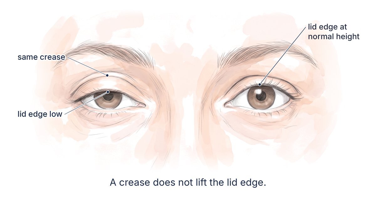
Ptosis correction and double eyelid surgery fix two different things. Double eyelid surgery makes a crease, the fold line above the lashes; ptosis correction, offered in Korean clinics as “eye-opening surgery” (nunmae gyojeong), raises a droopy lid edge (eyelid ptosis) so more of the iris and pupil show. A crease over mild ptosis still looks sleepy, so the first question is where your lid edge sits.
Quick answers
- Is my eye actually droopy? Measure MRD1, the gap from the upper lid edge to the light reflex in the pupil, with the forehead relaxed and any overhanging skin lifted. Normal is about 4 to 5 mm (3 to 3.5 mm on average in Asian adults); Dr. Yongwoo Lee recommends adding ptosis correction at 3.5 mm or less when the lid edge itself is low, not only covered by skin or a low brow.
- Will fixing one eye make the other droop? It can. After one-sided ptosis surgery from inside the lid in 75 patients, the other lid dropped later in 32 percent, enough to matter in 8 percent.
- Can it be done without an incision? For mild ptosis in a thin lid, Dr. Yongwoo Lee places a Müller’s muscle tuck through three small punctures.
Why Do My Eyes Still Look Sleepy After Double Eyelid Surgery?
Usually because the lid edge was already low before surgery, and a crease cannot lift it. A fold built over an undiagnosed mild ptosis sits on a lid that still covers the top of the iris, and the skin under the crease can bunch into a puffy roll.
In a series of 108 Asian patients, the authors note that about half of Asians lack a crease and many have a mild ptosis of 2 mm or less that surgeons may miss. A Korean five-year review of 127 patients put it plainly: performing non-incisional double eyelid surgery without correcting the ptosis tends to lead to unsatisfactory results.
A chapter on revision surgery lists ptosis among the reasons for unhappiness after a double eyelid operation: a lid with ptosis resists forming a fold, and ptosis can coexist with multiple creases. The puffy roll it leaves is one form of the “sausage eye” that needs a different revision from a fold set too high.
Hence the long-standing rule, cited in a 2024 prospective study of 97 Asian patients, that even a mild 1 to 2 mm ptosis should be corrected before any attempt to create a crease.
Is Ptosis Correction the Same as Double Eyelid Surgery?
No. Double eyelid surgery decides where the skin folds, and ptosis correction decides how high the lid edge rises. They are often done together through one incision, but they are separate steps with separate measurements.
Ptosis correction works one layer deeper than the crease, on the two muscles that open the lid.
The two lifters
The main lifter is the levator. Its broad tendon, the levator aponeurosis, attaches to the tarsal plate, the firm plate inside the lid. Behind it lies a smaller lifter, Müller’s muscle, which runs to the upper edge of the tarsal plate and raises the lid about 2.5 mm when it contracts. Eye-opening surgery shortens one or both of these so the lid rests higher.
What counts as mild ptosis
Ptosis is graded by how far the lid sits below where it should: mild 1 to 2 mm, moderate 3 to 4 mm and severe more than 4 mm. In a Korean national survey of 17,878 adults that counted ptosis as MRD1 under 2 mm, 12.08 percent met that definition, rising steeply with age.
Many cosmetic patients have a smaller droop than that. A 2024 prospective study of 97 Asian patients defines subclinical ptosis as MRD1 of 3.0 to 4.5 mm with good levator function, a heavy feeling and the forehead joining in to open the eyes.
How Do I Know If My Eyelid Is Actually Droopy?
Measure the lid edge instead of judging from the crease. Three checks decide it: MRD1, the distance from the light reflex in the pupil to the upper lid edge while you look at a light, normally 4 to 5 mm; levator function, taken with the brow held still; and a look at skin and brow for causes that only make the lid look low. At an MRD1 of 3.5 mm or less, Dr. Yongwoo Lee recommends correcting the lid along with the crease.
MRD1: the number that matters
In Asian adults MRD1 runs lower than the textbook 4 to 5 mm: one evaluation guide gives an average MRD1 of 3 to 3.5 mm in Asians, and suggests checking for lid retraction above 5.5 mm.

At home, with your forehead relaxed, look straight into a mirror with a phone light held at eye level beside it. A lid edge that reaches the top of the pupil or sits close to the light dot suggests ptosis. It is a starting point, not a measurement.
Levator function: how strong the lifter is
Levator function is the lid’s travel from full down-gaze to full up-gaze, with a finger fixing the brow so the forehead cannot help. It is typically 12 to 17 mm; good is 8 mm or more, fair 5 to 7 mm and poor 4 mm or less. Mild cosmetic ptosis usually has good function, so the lid can be tightened from its own muscles.
Frontalis compensation: the forehead that helps
When the lid is weak, the forehead muscle, the frontalis, lifts the brow to pull skin off the lashes. That overaction can cause brow fatigue and headache, and it leaves horizontal forehead lines even when the person is not trying to raise the brows.
After the lid is corrected, the forehead relaxes and the brow usually comes down, which can add heaviness if the brow was already low. Forehead toxin can also unmask a lid the frontalis was holding up, as in droopy eyelid after Botox.
Pseudoptosis: a lid that only looks low
Pseudoptosis means the lid edge sits normally but something makes it look low. Overhanging skin, a low brow, lid swelling, a sunken or downward-turned eye and a retracted lid on the other side can all do it.
Two simple tests separate it. One review advises that the upper eyelid skin should be manually elevated to see whether a true ptosis lies underneath. For Asian lids with MRD1 under 3.5 mm, the evaluation guide adds a crease test: if making a crease line with a thin wire creaser improves the lid height, it is likely pseudoptosis. In that lid a crease alone may be enough.
The other eye
Both levators receive one shared signal. Lifting one lid by hand can make the other descend, because in one-sided ptosis the better lid is held up by extra drive. That is Hering’s law, and lifting the droopier lid during the exam can reveal a hidden droop on the other side.
Which Operation Fixes Mild Ptosis: Müller’s Tuck, MMCR or Levator Plication?
A crease operation, by buried suture or through an incision, treats the fold. A low lid edge is raised by tightening Müller’s muscle, folding or advancing the levator, or, when the levator barely works, hanging the lid from the forehead muscle with a sling.
Müller’s muscle tuck by suture
A suture folds Müller’s muscle on itself, removing nothing. In one Korean series of incisional double eyelid surgery with this tuck, 1 mm and 2 mm of correction could be achieved predictably with one and two loops, though in thick, scarred lids the correction could fall by as much as 50 percent.
It can also be placed without an incision. In 127 patients having a non-incisional fold with a Müller’s tuck averaging 7.8 mm, MRD1 rose from 1.62 mm to 3.97 mm. In a series of 161 patients having a transconjunctival tuck, only 34 were followed beyond six months; in them MRD1 went from 2.08 mm to 3.49 mm right after surgery and 3.22 mm at six months, with 3 of 34 patients re-corrected. These are retrospective case series, not trials.
Müller’s muscle and conjunctival resection
MMCR works from inside the lid and removes a strip of Müller’s muscle with its lining. It classically suits about 2 to 3 mm of ptosis that responds to phenylephrine drops, with levator function over 10 mm, and leaves no skin scar. A pooled analysis of 7 studies and 1,038 eyelids found similar final lid height to levator advancement, with less overcorrection and fewer reoperations.
In one retrospective series of 404 lids, success was 90 percent after MMCR and 79 percent after levator surgery. Part of that gap is selection, since the inside approach is usually chosen for smaller droops.
Levator plication or advancement
Through the crease, the levator can be folded on itself (plication) or detached and advanced. In 108 Asian patients having a double eyelid with levator aponeurosis plication, MRD1 rose from 2.93 mm to 4.21 mm, with 3.2 percent undercorrected and 1.3 percent overcorrected.

Frontalis sling
When levator function is under about 5 mm, the lid cannot be tightened from its own muscles, and it is hung from the forehead muscle with a sling.
At a Glance: Crease Surgery vs Ptosis Correction
| Option | What it changes | Access | Typically suited to | What the evidence shows |
|---|---|---|---|---|
| Double eyelid surgery alone | Crease only | Punctures or crease incision | MRD1 in range, no forehead help | Does not raise the lid edge |
| Müller’s tuck through the crease (Dr. Yongwoo Lee’s first choice) | Lid height, then crease | Crease incision | Mild ptosis, good levator function | Korean series: about 1 mm per loop; no published data for his exact method |
| Müller’s tuck through punctures | Lid height with a stitched fold | Three punctures | Mild ptosis in a thin lid, little excess skin or fat | Related transconjunctival tuck series: MRD1 2.08 to 3.22 mm at six months in the 34 of 161 patients followed |
| Under-through levator plication (Dr. Yongwoo Lee) | Lid height, more lift | Crease incision | Tuck not enough, a little more ptosis, revisions | Related plication series: MRD1 2.93 to 4.21 mm |
| MMCR | Lid height | Inside the lid | 2 to 3 mm, drop-responsive | 90 vs 79 percent success against levator surgery, one series |
| Levator advancement | Lid height | Crease incision | Moderate ptosis | More overcorrection and reoperation in pooled studies |
| Frontalis sling | Lid lifted by forehead | Lid and brow | Levator function under 5 mm | Referred to a ptosis specialist |
How Is Mild Ptosis Corrected During Double Eyelid Surgery? Dr. Yongwoo Lee’s Method
When MRD1 is 3.5 mm or less, I recommend correcting the ptosis in the same operation, through the same crease incision, before the crease is fixed. My first choice is a Müller’s muscle tuck at three points; if it does not lift enough with the patient awake, I switch to an under-through levator plication.
Where I draw the line
That threshold sits above the 2 mm line of population surveys, which count lids low enough to be a medical finding; a cosmetic plan asks whether the eye will look right under a new crease. Before I mark anything, I measure MRD1 with the brow relaxed and levator function with the brow held still, and I lift the brow with a fingertip to see how much of the heaviness is brow rather than lid. I count only a lid edge that stays low with the brow relaxed and the excess skin lifted, and I exclude pseudoptosis.
Three reference points set by the iris
I set three reference points on the lid by the iris: over its center, its inner edge and its outer edge. Every stitch that changes lid height goes in at these three points.
The Müller’s muscle tuck, stitch by stitch
I use 7-0 blue nylon. At each point the needle enters the tarsal plate about 2 mm below its upper border and passes from front to back, to the inner lining of the lid, the conjunctiva. It re-enters the same hole and runs up beneath the conjunctiva for about 7.5 mm, catches Müller’s muscle, runs back down beneath the conjunctiva, and comes back out through the tarsal plate.
I tie it on the front of the tarsal plate. The knot sits in front, inside the wound, so it does not lie against the eye. In mild ptosis the tuck is usually 6 to 9 mm, often about 7.5 mm, but it varies with the lid and is sometimes more.
When the tuck is not enough: under-through levator plication
If the tuck does not lift enough when you open your eyes, I switch to an under-through levator plication. I also use it from the start when the ptosis is a little more than mild, and in revision cases, where scar can blunt a tuck.
Müller’s muscle is not separated from the levator. At each of the same three points I place one stitch. It takes a partial-thickness bite of the tarsal plate about 2 mm below its upper border, runs up on the underside of the levator aponeurosis for about 7.5 mm, passes through the aponeurosis and is tied on its front. As it tightens, the aponeurosis and Müller’s muscle fold together as one complex, with Müller’s muscle meeting Müller’s muscle inside the fold, so the two surfaces heal together fairly evenly and the lift is more predictable. The fold sits on the front of the tarsal plate, and the levator is not detached from it.

Without an incision: three punctures
When the eyelid is thin, with little excess skin or fat, I place the Müller’s tuck through three small punctures instead of a crease incision and make the fold with a buried suture in the same operation. More ptosis, a thick lid or a revision goes to the incisional route.
Why I wake you, several times
I give the anesthesia myself: IV sedation with midazolam and ketamine plus local anesthesia, light enough to wake you, while a dedicated nurse watches the monitors for blood pressure, oxygen level and heart rate throughout. During the operation I wake you and ask you to open your eyes, then check lid height and symmetry, and I do it several times.
The reason is Hering’s law: raising one lid can let the other drop slightly. It does not happen in everyone, and in one study a drop test before surgery did not predict who it would happen to. Each time you open your eyes, I add to or reduce the tuck. If it is clearly too much or too little, I untie it and place it again.
Before raising any lid, I ask about dry eye and check that both eyes close completely, because a higher lid exposes more of the eye’s surface.
The crease comes last: TAO fixation
Only when the height is right do I make the crease. I use TAO fixation (tarsus, aponeurosis, orbicularis): a partial bite of the tarsal plate near its upper edge, the levator aponeurosis, the orbicularis muscle and the skin edge are joined, so the fold moves with the lid. I always keep the crease within the height of the tarsal plate. Why the tarsal plate sets the ceiling, and how buried-suture and incisional folds differ, is covered separately.
What I do not take on
Severe ptosis, and a lid with poor levator function that needs a sling, I refer to a ptosis specialist.
Video in English, about 12 minutes: Upper Blepharoplasty & Mild Ptosis Correction in 3D | Müller’s Tuck & Under-Through Plication
Do You Need Ptosis Correction, a Double Eyelid or Both? Six Eyelid Profiles
Two findings decide it: whether the lid edge is low when measured, and whether the eyelid is thin enough for a stitched fold. These illustrative examples, not real patients, describe decisions, not results.
No crease, MRD1 in range, quiet forehead. A crease alone. A thin lid can have a buried-suture fold, and how long a buried-suture fold lasts depends mostly on the lid it is placed in.
No crease, sleepy look, MRD1 3.5 mm or less, forehead lines when you open your eyes. Crease plus a Müller’s tuck through the same incision: the typical hidden mild ptosis.
Thin lid, mild ptosis, wants no incision. A buried-suture fold with a Müller’s tuck through three punctures, if the lid has little excess skin or fat.
A double eyelid that still looks sleepy, with a puffy strip under the crease. If MRD1 confirms the lid edge is low, revision with an under-through levator plication from the start, after the earlier operation has settled; if the lid height is normal, the fold itself is the problem, as in a sausage eye.
One eye clearly smaller. Both lids are measured, and you are warned beforehand that the other may come down once the first is lifted. A new or fluctuating one-sided droop is checked for a nerve or muscle cause first.
Looks droopy, but MRD1 is normal when skin or brow is lifted. Pseudoptosis: skin removal, a crease or brow work, not ptosis surgery. If the heaviness sits under the brow, a sub-brow lift may fit better than a lid operation.
What Is Recovery Like After Ptosis Correction With Double Eyelid Surgery?
Swelling and bruising are heaviest in the first days and largely fade over about two weeks, and skin stitches come out on day 7 after an incisional operation, counting the day after surgery as day 1. Lid height keeps settling for weeks, so it is judged at about three months.
Dressings are changed almost daily at first. Mild incomplete closure usually resolves within 2 to 3 weeks. In 92 older patients, temporary incomplete closure occurred in every ptosis repair and mostly resolved within 3 months, with dry-eye scores worst at one month. Lubricating drops by day and an ointment at night, while the eye does not close fully, are the usual support in that window, and dry eye and incomplete closure after eyelid surgery have their own article.
Travelers should plan to stay at least until the stitches are out; see how long to stay in Korea after surgery.
Surgeon’s Insight: A Crease Cannot Open an Eye
Patients bring photographs of folds. What the photograph also shows, and what they rarely notice, is how much of the iris the lid leaves uncovered. Put the same fold on a lid that sits a millimeter or two low, and it reads as tired.
So I measure the lid edge before I discuss the fold, set the height before I fix the crease, and check both eyes open before I finish, because the other eye has a vote.
What Are the Risks of Ptosis Correction, and What Should I Watch For?
The commonest problems are a lid left slightly low and asymmetry; early incomplete closure and dry eye are common and usually temporary. Less common are overcorrection, an uneven lid contour, the other lid dropping, and slow recurrence over years.
Undercorrection is the most frequent complication of ptosis surgery, seen in about 10 to 15 percent, and some apparent undercorrection is swelling that settles. In a single-surgeon series of 2,328 patients at a Korean tertiary center, mostly congenital and more severe cases, reoperation by three years was 10.4 percent for acquired ptosis.
The other lid
After one-sided MMCR in 75 patients, the untreated lid dropped later in 32.0 percent, and enough to matter in 8.0 percent. Earlier reports cited by another group give contralateral droop in 9.6 to 17 percent after one-sided repair, more often after levator advancement than after MMCR. An evaluation guide adds that when the droopy eye is the dominant one, the other eye may be working harder and can drop once the first is corrected, so eye dominance should be checked first.

Overcorrection, lashes and symmetry
A lid raised too far exposes the eye and looks staring. Excessive correction can also turn the lashes upward and expose the inner lining of the lid. One review advises favoring a slight undercorrection when dry eye is severe or the lid may not close. Perfect symmetry is not certain: in congenital ptosis with a crease made at the same time, only 30 percent were perfectly symmetric, and asymmetry appeared by six months in many that first looked even.
Recurrence
In 404 lids, the risk factors for failure included prostaglandin eye drops, used for glaucoma, as the highest known risk, followed by topical steroids, the approach, a lower starting MRD1 and earlier surgery inside the eye, such as cataract surgery. Mention any long-term eye drops at consultation.
When to call
Rapidly increasing pain, a tense or bulging eye or any loss of vision after eyelid surgery is an emergency: call your local emergency number or go to the nearest emergency department first, then contact the clinic. A red, painful, light-sensitive or increasingly gritty eye, or blur that does not clear with blinking, can mean the eye’s surface is drying or scratched: contact the clinic the same day. A droop that appears suddenly, gets worse through the day or when you are tired, or comes with double vision or a pupil change, is not a cosmetic question; the warning signs are listed in droopy upper eyelids after 50.
What Should I Ask Before Choosing Double Eyelid Surgery or Ptosis Correction?
Ask for your MRD1 on each side, your levator function, and whether your forehead is helping you open your eyes. Those three answers decide whether you need a crease, a lid correction or both.
Ask, too, how the other eye was checked and whether you will be woken to open your eyes.
If you ask only one thing, ask this: Is my lid edge low, or is it only covered?
Individual results vary; consultation required.
Written by Dr. Yongwoo Lee, board-certified Korean plastic surgery specialist in facial anatomy and aesthetic procedures at VIP Plastic Surgery, South Korea.
Frequently Asked Questions About Ptosis Correction and Double Eyelid Surgery
How do I know if I have ptosis or just a hooded eyelid?
Lift the skin fold gently off the lashes, or lift the brow with a fingertip, and look at the lid edge. If it sits at a normal height, the lid is hooded; if it still covers the top of the pupil, ptosis is likely. Measurement confirms it.
What is a normal MRD1, and what counts as mild ptosis?
Normal MRD1 is about 4 to 5 mm, with an Asian average of 3 to 3.5 mm, and a lid 1 to 2 mm below normal is graded mild ptosis. A cosmetic plan sets its own line: Dr. Yongwoo Lee adds ptosis correction at 3.5 mm or less, measured with the forehead relaxed.
Is eye-opening surgery the same as ptosis correction?
In most Korean clinics, eye-opening surgery (nunmae gyojeong) means correction of mild ptosis, often combined with a double eyelid. The term is used loosely, so ask which muscle is being tightened.
Can I get a double eyelid without fixing mild ptosis?
You can if the lid edge sits at a normal height, or if it only looks low and a crease itself lifts it (pseudoptosis). Over a truly low lid, a crease tends to sit puffy over an eye that still looks sleepy.
Does non-incisional ptosis correction last?
It can, in a thin lid, but follow-up is short: in the one series followed past six months, most of the lift held and 3 of 34 patients were re-corrected. Thick or scarred lids suit the incisional route better, and the fold made with it is a buried-suture fold, with that method’s own durability limits.
What is the difference between a Müller’s muscle tuck and levator plication?
A Müller’s tuck folds the smaller lifting muscle with a stitch and suits mild ptosis. Levator plication folds the main lifting tendon and gives more lift. Dr. Yongwoo Lee starts with the tuck and, if it falls short or in revisions, folds both together in an under-through plication.
Will ptosis correction make my eyes look bigger?
It shows more of the iris and pupil, so the eye looks more open and alert. It does not widen the eye from corner to corner.
Will fixing one droopy eyelid make the other one droop?
It can, because both lids receive one shared signal to open (Hering’s law), and in one study a drop test before surgery did not predict who would be affected. That is why both lids are checked open during surgery, and why eye dominance is checked first when the droopier eye is the dominant one.
Will I be able to close my eyes, and will they feel dry?
A small gap on closing is common for the first few weeks and usually resolves; in older patients, dry-eye symptoms were worst at one month and mostly settled by three to six months. Mention any dry eye at consultation.
What if the lid ends up too high or too low?
Some apparent undercorrection is swelling, so lid height is judged at about three months, when a lid still low is assessed for adjustment; a crease problem is given at least six months. A lid too high to close is seen sooner.
Is congenital ptosis treated differently?
Often, yes. In a Korean series of 2,328 ptosis patients, mostly congenital, a forehead sling was the commonest operation (55.1 percent), then levator resection (29.0 percent). Severe ptosis and poor levator function are referred to a ptosis specialist.
Am I awake during ptosis correction?
Partly. Dr. Yongwoo Lee gives IV sedation with midazolam and ketamine plus local anesthesia himself, light enough to wake you several times to open your eyes, so height and symmetry can be adjusted before the crease is fixed.
This article is for informational purposes only and does not constitute medical advice. Always consult with a qualified medical professional before making any decisions about surgical or non-surgical procedures.
